


Chromosomes are structures within cells that contain a person's genes.
Genes are segments of deoxyribonucleic acid (DNA) and contain the code for a specific protein that functions in one or more types of cells in the body (see Genes and Chromosomes for a discussion about genetics).
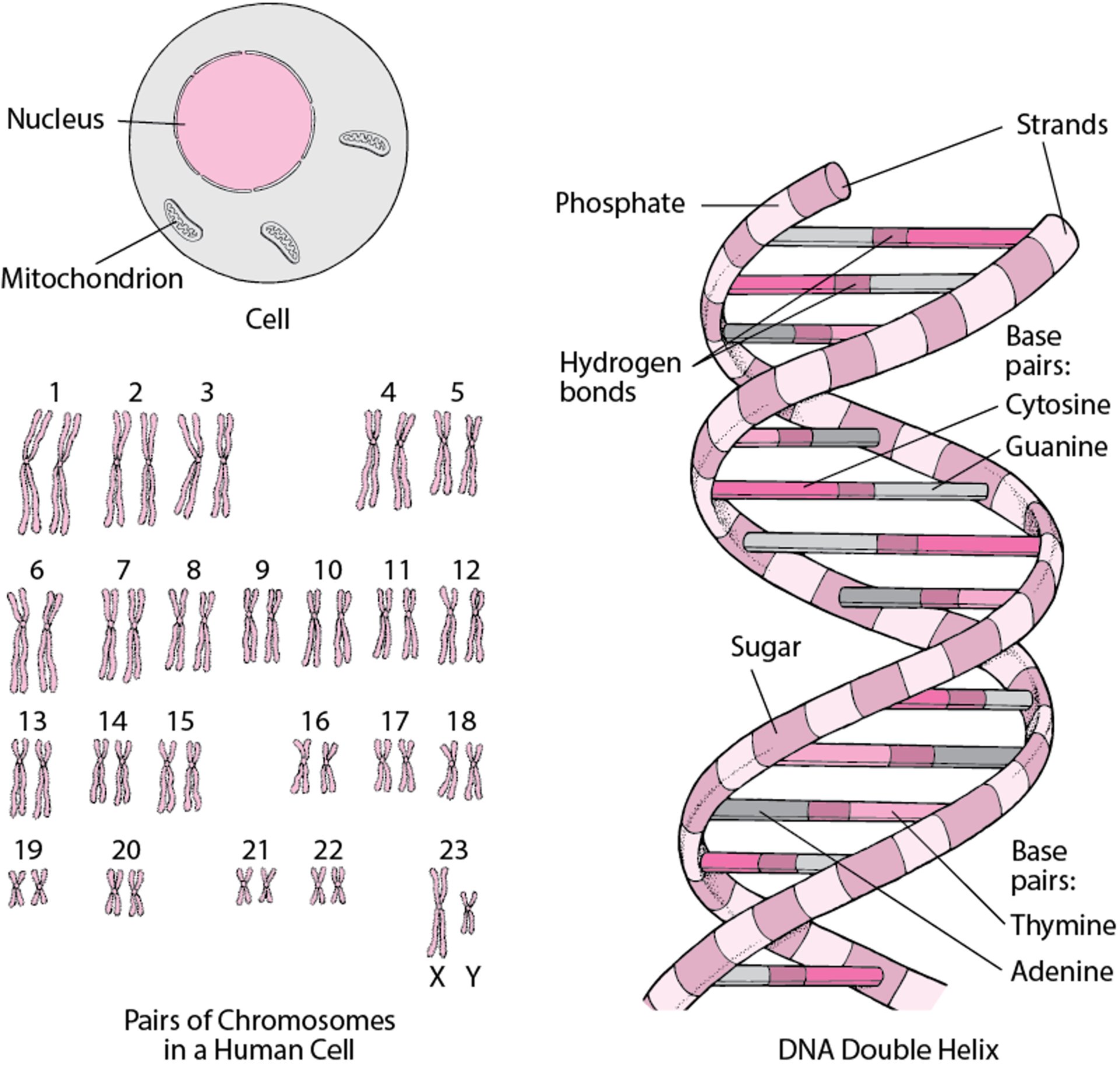
Every normal human cell, except for sperm and egg cells, has 23 pairs of chromosomes for a total of 46 chromosomes. Sperm and egg cells have only one of each pair of chromosomes for a total of 23. Each chromosome contains hundreds to thousands of genes.
The sex chromosomes are one of the 23 pairs of chromosomes. There are 2 sex chromosomes, called X and Y. Females typically have two X chromosomes (XX) and males typically have one X chromosome and one Y chromosome (XY).
Structure of DNA
DNA (deoxyribonucleic acid) is the cell’s genetic material, contained in chromosomes within the cell nucleus and mitochondria. Except for certain cells (for example, sperm and egg cells and red blood cells), the cell nucleus contains 23 pairs of chromosomes. A chromosome contains many genes. A gene is a segment of DNA that provides the code to construct a protein or RNA molecule. The DNA molecule is a long, coiled double helix that resembles a spiral staircase. In it, two strands, composed of sugar (deoxyribose) and phosphate molecules, are connected by pairs of four molecules called bases, which form the steps of the staircase. In the steps, adenine is paired with thymine and guanine is paired with cytosine. Each pair of bases is held together by a hydrogen bond. A gene consists of a sequence of bases. Sequences of three bases code for an amino acid (amino acids are the building blocks of proteins) or other information. |

Chromosome Abnormalities
(See also Chromosomal abnormalities under Risk Factors for Genetic Disorders or Birth Defects.)
Chromosome abnormalities can affect any chromosome, including the sex chromosomes. Chromosome abnormalities affect the
Number of chromosomes
Structure of chromosomes
Larger abnormalities may be visible with a microscope in a test called chromosome analysis or karyotyping. Smaller chromosome abnormalities can be identified using specialized genetic tests that scan a person's chromosomes for extra or missing parts. These tests include chromosomal microarray analysis (CMA) and fluorescent in situ hybridization (FISH). (See also Next-generation sequencing technologies.)
Numerical abnormalities occur when a person has one or more extra copies of a chromosome (for example, one extra is trisomy, and two extra is tetrasomy) or is missing an entire chromosome (monosomy) or part of a chromosome. Trisomy can affect any of the 23 paired chromosomes, but the most common are trisomy 21 (Down syndrome), trisomy 13, and trisomy 18, which affect both boys and girls. These abnormalities are visible with a microscope in karyotyping.
The older a pregnant woman is, the greater the chance that her fetus will have a whole extra chromosome or will be missing a chromosome (see table What is the Risk of Having a Baby With a Chromosomal Abnormality*?). The same is not true of a man. As a man gets older, the chance of conceiving a baby with a chromosome abnormality is only slightly increased.
Structural abnormalities occur when part of a chromosome is abnormal. Sometimes part or all of a chromosome incorrectly joins with another chromosome (called translocation). Sometimes parts of chromosomes are missing (called deletion―see Overview of Chromosomal Deletion Syndromes) or have been duplicated.
Some chromosome abnormalities cause the death of the embryo or fetus before birth. Other abnormalities cause problems such as intellectual disability, short stature, seizures, heart problems, or a cleft palate.
Gene Abnormalities
Changes in one or more base pairs of the DNA in a gene (see figure Structure of DNA create a variant of that gene that may affect how the gene works. These changes do not affect the structure of the chromosomes and thus cannot be seen on karyotype analysis or other chromosomal tests. More specific genetic testing is required. Some variants in a gene cause no problems and some cause few or only mild problems. Other variants cause serious disorders such as sickle cell anemia, cystic fibrosis, and muscular dystrophy. Increasingly, medical scientists are finding specific genetic causes of children's diseases.
It remains unclear how most variants occur, but most are thought to appear spontaneously. Some substances or agents in the environment are capable of damaging and causing variation in genes. These substances are called mutagens. Mutagens, such as radiation, ultraviolet light, and certain medications and chemicals, can cause some cancers and birth defects.
A variant in the genes in a sperm or egg can be passed from parent to child. Variants of genes in other cells may cause disease that is not passed down to children (because the sperm or eggs are not affected). Having two copies of an abnormal gene can lead to serious diseases or conditions, such as cystic fibrosis or Tay-Sachs disease. Sometimes disorders can occur even when a person has just one copy of an abnormal gene.
Testing for Chromosome and Gene Abnormalities
A person's chromosomes and genes can be evaluated by analyzing a sample of blood as well as cells from other parts of the body such as from a swab of the inside of the cheek.
During pregnancy, doctors can use cells retrieved via amniocentesis or chorionic villus sampling to detect certain chromosome or gene abnormalities in a fetus. If the fetus has an abnormality, further tests may be done to detect specific birth defects.
More recently, a screening test has been developed in which a sample of a pregnant woman's blood is analyzed to determine whether her fetus has an increased risk of certain genetic disorders. This test is based on the fact that the mother's blood contains a very small amount of DNA from the fetus. This test is called noninvasive prenatal screening (NIPS) or cell-free fetal DNA analysis. NIPS can be used to detect an increased risk of trisomy 21 (Down syndrome), trisomy 13, or trisomy 18 and certain other chromosome and gene disorders but is not diagnostic. Doctors usually recommend further testing when any chromosome abnormality is detected.
Prevention of Chromosome and Gene Abnormalities
neural tube defects, and parents can be screened for carrier status of certain genetic abnormalities. An embryo conceived through in vitro (test tube) fertilization (IVF) can also be tested for a genetic abnormality before it is transferred into the woman’s uterus (see Preimplantation Genetic Diagnosis).


